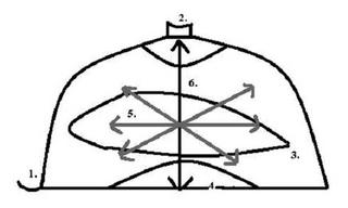
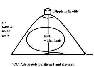
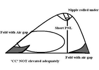
WHAT MUST WE SEE IN THE CC PROJECTION?
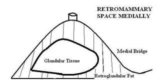
- MEDIAL NUB
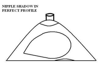
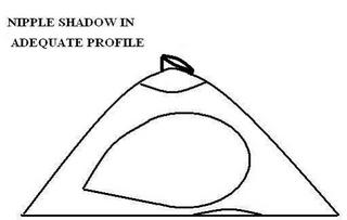
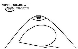
- NIPPLE IN PROFILE
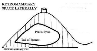
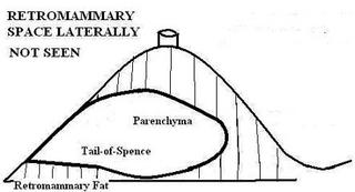
- TAIL OF SPENCE
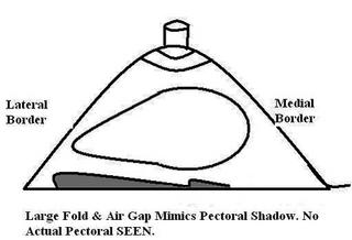
- PECTORAL SHADOW
- TISSUE SPREAD ADEQUATELY
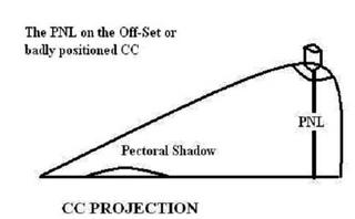
- PNL WITHIN 1CM OF MLO
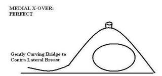
WHEN DO WE SEE THE MEDIAL CROSS-OVER?
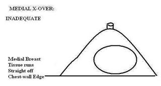
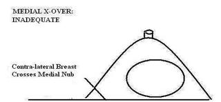
WHEN DON'T WE SEE THE MEDIAL CROSS-OVER?
WHEN DON'T WE SEE THE NIPPLE IN PROFILE?
WHEN DO WE SEE RETRO MAMMARY SPACE?
Adequately Demonstrated Pectoralis
WHEN DON'T WE SEE PECTORAL SHADOW?

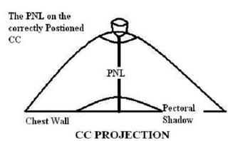
MEASURING THE POSTERIOR NIPPLE LINE
The PNL on the CC projection is ALWAYS measured from the base of the nipple directly back to the film edge. The measurement is taken this way irregardless of pectoralis shadow or improper positioning.
Our mission as mammographers gets more complicated every year. We are called upon to assess patients, images, equipment and services. Breast imaging is complex but not beyond skills. Everything follows certain recognized standards, adhere to these and things will not seem so demanding.
