The PNL dimension must be within 1cm of measured distance on both the CC and the MLO projection. If the PNL is much shorter on one view than the other, there is inadequate tissue on that projection.
Measuring the PNL on the CC projection:
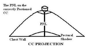
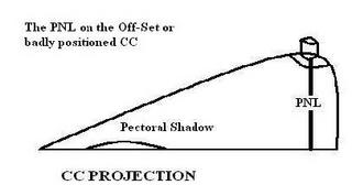
The posterior nipple line in the cranial-caudal projection is measured from the nipple base junction directly to the back of the image. The PNL on the CC is always calculated to the very back of the film edge regardless of the image of the pectoral shadow. The PNL on the CC image MUST be within 1cc of the PNL on the MLO view. Obviously, these measurements loose validity if your MLO was positioned poorly. However, the PNL still provides a simple mechanism for comparative assessment of the adequate depth of the CC projection.
THE PNL ON THE CC:

 Measuring the PNL on the MLO projection:
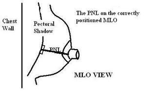
The Posterior Nipple Line on the MLO view should be measured from just behind the nipple base directly parallel to the cooper’s ligaments to the front of the pectoral shadow or to the film edge, which ever comes first. The PNL on MLO should be gauged by placing your straight edge 90˚ to the edge of the pectoral shadow and sliding it down to the base of the nipple. The PNL of the MLO must measure within 1cm of the PNL on the CC.
MEASURING THE PNL ON THE MLO:
Sometimes, it is true that a picture is worth 1000 words. Describing the PNL is difficult and often confusing but it is an excellent device for us to ensure our breast images are accurate, complete and diagnostic. The PNL measurement should not become an embarrassing test for us; it should be a very useful tool.
The criteria used for the PNL span is established in articles published by Kopans, Eklund and Cardivosa. These conventions for measuring this parameter are used in the facility accreditation process and in the technologist’s certification procedure.


